
Partial Parallel Imaging (PPI) Brain Scans
jpg’s of the 3 images:
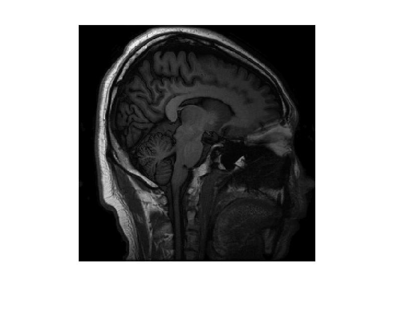
- Image 1
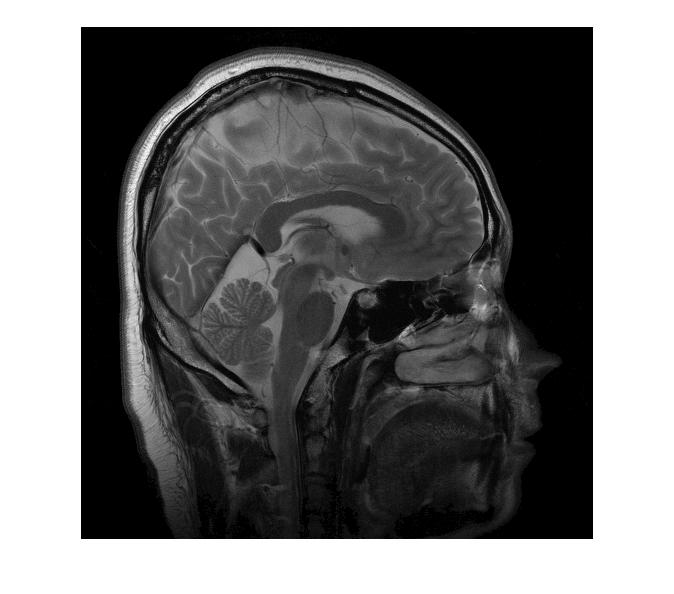
- Image 2
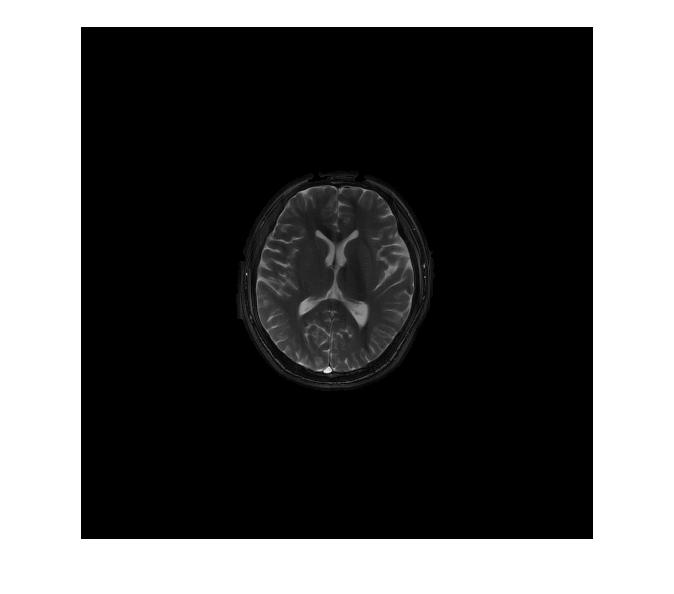
- Image 3
The 8-channel PPI data for each image is stored inside a MATLAB “mat” file. Each file contain 3 arrays: p (a mask, either random or radial), sense_map (sensitivity map), and u0 (image). The image can be viewed in MATLAB with the command imshow(u0). The mask can be viewed with the low frequency at the center with the command imshow(fftshift(p)). The data corresponds to equation (1) in the paper “Fast Algorithms for Image Reconstruction with Application to Partially Parallel MR Imaging.” u0 in the data file corresponds to u in equation (1). The output of the j-th receiver is the product M F S_j u where u is the image, M is the mask, F is the Fourier transform, and S_j is the sensitivity map.
- data 1
- data 2
- data 3
- June 9, 2013.