
Research
Using behavioral, physiological, and brain imaging methods, Dr. Keil and his colleagues study how our perception of the physical world changes depending on our internal states and goals and how it is shaped by experience. Specifically, we are interested in how sensory systems in the brain support mental health, how they indicate brain dysfunction, and how we can use the plasticity of sensory systems in the human brain to promote well-being. At a methodological level, we are interested in developing and validating methods to quantify human brain activity. Together with our colleagues at the University of Florida College of Engineering, we are conducting simultaneous EEG-fMRI recordings, exploring the temporal and spatial dynamics of the human brain during tasks that challenge perception, attention, and emotion. Our laboratory collaborates with numerous researchers at UF, including in the colleges of Medicine, Education, and Engineering, and with many laboratories across the globe.
Experience and visual perception
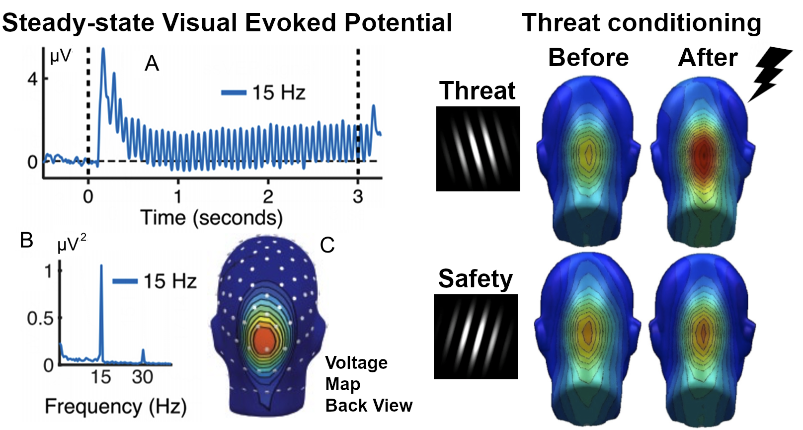
Traces of experiences, good and bad, are stored throughout the brain and body. We study the way visual cortical neurons adapt to environmental challenge, implemented for instance as fear conditioning in the laboratory.
Distraction and competition
Our perceptual systems have limited capacity but are constantly exposed to a barrage of information conveyed by the senses. In a constantly changing and complex visual world, attention is needed to focus on relevant information while ignoring other things This is often a challenge, especially over longer periods of time (e.g., Wieser & Keil, 2011). For instance, one may try to do homework but become distracted by a cartoon advertisement on a website or by the stock ticker scrolling on bottom of the screen. Engaging, emotionally arousing stimuli are particularly powerful distractors. We examine the extent, time course, and cortical mechanisms of distraction. An example paradigm used in our laboratory to assess competition effects between different stimuli over time is the attentional blink task (e.g., Keil & Ihssen, 2004; Heim & Keil, 2012).

Oscillatory Brain Activity and Behavior.
The human brain is one of the most complex systems known. Although no comprehensive theory of brain function exists, there is an emerging consensus that it is best conceived as a flexible and dynamic, massively parallel system, in which behavior and cognition emerge through oscillatory communication among units and subsystems. We study oscillatory brain dynamics using dense-array electrophysiology and simultaneous EEG-fMRI recordings in humans (e.g. Liu et al. 2012) and develop methods to better quantify and characterize oscillatory brain dynamics in the context of perception, emotion, and learning (e.g., Miskovic & Keil, 2014). Method development greatlt benefits from our ongoing collaborations with Drs. Ding and Principe, UF College of Engineering.
Psychophysiology of emotion, fear, and anxiety: Interindividual differences.
Many psychiatric disorders are characterized by dysregulation of emotional reactivity, for instance when patients are exposed to potentially threatening situations. Using measures of brain function, autonomic ractivity, behaviroral responses, and self report, we examine how biomarkers of emotional processing in the laboratory can be used to characterize self-reported problems associated with disorders of mood and anxiety (e.g., Moratti et al., 2008). Establishing reliable and valid quantitative indices of inter-individual differences in affective reactivity is a necessary step towards objective assessment of the brain circuits underlying emotional dysfunction in anxiety and mood disorders, and ultimately to inform evidence-based intervention and treatment efforts.
