
Areas of Research
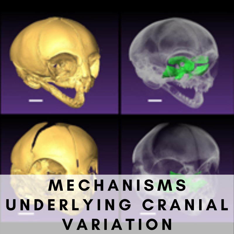
Dr. Valerie DeLeon and the members of the DeLeon Lab use 3D virtual morphology to investigate sources of anatomical variation in humans and other primates.


We generate 3D PDFs to let our audience interactively view important structures in three dimensions.
Image: Sphenoid of Callicebus


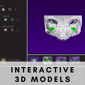
Interactive 3D Models
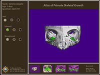
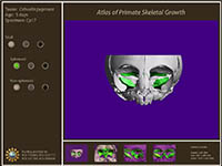
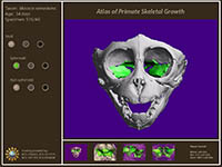
Sphenoethmoidal Junction in Perinatal Primates
 |
 |
 |
||
| Varecia variegata (31 MB) | Cebuella pygmaea (32 MB) | Macaca nemestrina (55 MB) |
These modules highlighting comparative anatomy of the sphenoethmoid junction were produced in our collaborative work with Dr. Timothy Smith, Slippery Rock University. These modules are interactive 3D PDF files that allow visualization of the sphenoid in three perinatal primates: a strepsirrhine (Varecia variegata), a platyrrhine (Cebuella pygmaea), and a catarrhine (Macaca nemestrina).
Click the thumbnails to open each interactive module in a new window.
[Note: Interactive modules require Acrobat Reader and JavaScript plugins for your internet browser. You can also download the file by right-clicking and saving the PDF to your computer. The large file size of these PDFs may result in delays depending on your internet connection.]
Elements of the Varecia and Cebuella modules were extracted for our 2017 paper in the Anatomical Record:
Smith, T.D., McMahon, M.J., Millen, M.E., Llera, C., Engel, S.M., Li, L., Bhatnagar, K.P., Burrows, A.M., Zumpano, M.P. and DeLeon, V.B., 2017. Growth and development at the sphenoethmoidal junction in perinatal primates. The Anatomical Record. DOI: 10.1002/ar.23630 (LINK).
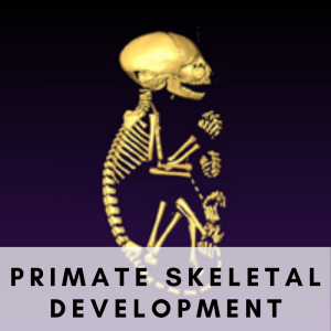
Primate Skeletal Development
Hyoid Growth in rhesus macaques
Posted on August 12, 2020 in News

Check out Andree’s infographic on her recent publication “Growth and sexual dimorphism of the hyoid body in Macaca mulatta” in the International Journal of Primatology.
Read her article here: https://rdcu.be/b4Tr6

Click on Andree’s profile to learn more about her research!